
Thoracentesis
Breathe easier by draining fluid around the lung.
Thoracentesis removes fluid that has built up around the lung, using a thin tube placed between the ribs under ultrasound. It takes 15 to 30 minutes, you stay awake, and many people breathe easier right away.
- Outpatient
- 15–30 minutes
- Numbing medicine
- Awake throughout
 Fabio Komlos, MDInterventional radiologist
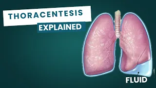 Watch the video explanation12:23 video · Thoracentesis Procedure: Everything You Need to Know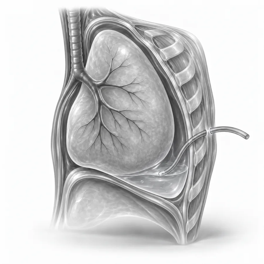
Numbing medicine and a thin, soft tube.
Thoracentesis Procedure: Everything You Need to Know
Dr. Fabio Komlos walks through thoracentesis the way he explains it in clinic, using the same images he uses to plan treatment.
Fabio Komlos, MDInterventional radiologist · 12:23PleurX Catheter
8:23
What is a pleural effusion?
Your lungs are wrapped in two thin, slippery layers called the pleura. Normally there are only a few teaspoons of fluid between them. When too much fluid builds up in this space, it is called a pleural effusion. It can happen on one side or both.
The fluid takes up room the lung needs to expand. A large effusion can squeeze part of the lung flat. You may feel short of breath, especially when walking or lying flat, have a dry cough or feel chest heaviness.
Common causes
- Heart failure, the most common cause
- Pneumonia and other chest infections
- Cancer, either in the lung or spread from elsewhere
- Liver cirrhosis or kidney disease
- Blood clots in the lung, recent surgery or inflammation
On ultrasound, fluid around the lung looks black. The lung floats in it as a gray, moving shape, and the diaphragm is a bright curved line below. We choose a spot where the fluid pocket is deepest, well away from the lung, liver or spleen.
Why thoracentesis helps
Thoracentesis does two jobs. Therapeutic drainage removes fluid so you can breathe more easily. Diagnostic drainage sends a sample to the lab to find the cause. Often we do both at once. We may also draw a blood sample to compare with the fluid.
Lab tests check protein, sugar, cell counts, signs of infection and cancer cells. These tell your doctor whether the fluid is a leak caused by pressure or low protein, as in heart failure (a transudate), or a sign of inflammation, infection or cancer (an exudate).
Good candidates
- A new effusion with no known cause
- A large effusion causing shortness of breath
- Suspected infection in the fluid
- A known effusion that has come back
May need a different plan if
- The fluid pocket is very small or split into compartments
- You have a bleeding problem that cannot be corrected
- The fluid returns quickly after every drainage
- The fluid is thick and infected, which often needs a chest tube
If the fluid keeps coming back
Treating the cause, for example with heart failure medicines or antibiotics, often stops the fluid from returning. If it keeps coming back, especially with cancer, there are longer-term options.
| Repeat thoracentesis | Tunneled pleural catheter | Pleurodesis | |
|---|---|---|---|
| What it is | Drain the fluid again as needed | A soft tube under the skin, drained at home | Seals the pleural space so fluid cannot collect |
| Best for | Fluid that returns slowly | Fluid that returns fast, or a lung that cannot fully expand | Fluid that returns, with a lung that fully expands |
| Visits | A new procedure each time | One procedure, then home drainage | One procedure, often a short hospital stay |
Read more about chest PleurX catheters and pleurodesis.
How to prepare
- 1Tell us about any blood thinners. Many people can keep taking them for thoracentesis, but we will confirm. See blood thinners.
- 2You can usually eat and drink normally, because no sedation is needed.
- 3Take your usual medicines, including water pills and inhalers.
- 4Bring someone with you if you feel very short of breath, but most people can drive themselves home.
What happens during thoracentesis
- 1Sitting up
You sit on the edge of the table and lean forward, resting your arms on a padded stand. If you cannot sit, we can do it lying on your side.
- 2Ultrasound and numbing
We find the fluid with ultrasound, mark the spot on your back, clean the skin and numb it down to the pleura. You feel a sting and then pressure.
- 3Draining the fluid
A thin needle with a soft plastic tube over it goes between two ribs into the fluid. The needle comes out, leaving only the soft tube, and fluid drains into a vacuum bottle.
- 4Finishing up
We remove the tube, place skin glue or a bandage, and send samples to the lab.
As the lung re-expands, it is common to feel chest tightness or the urge to cough, usually near the end. Tell us, because it often means it is time to stop. We usually remove no more than about 1 to 1.5 liters at a time to protect the lung.
I always scan the chest right before I start, even if you had a scan yesterday, because fluid shifts. I look for the deepest pocket with a safe margin from the lung. I also watch you as much as the ultrasound: a cough or tightness tells me the lung is coming back up, and that is usually when I stop.Fabio Komlos, MD, interventional radiologist · About Dr. Fabio Komlos
Recovery
- Right afterRest for a short while. Some people have a chest X-ray, especially if they have new symptoms. Many breathe more easily right away.
- Same dayYou can eat normally and shower. Most people can drive home.
- First 24 hoursAvoid heavy lifting and hard exercise. Mild soreness at the site is normal.
- Day 2 onwardBack to your usual activities. Your doctor will call with lab results, usually within a few days.
An ice pack and acetaminophen (Tylenol) or ibuprofen usually control any soreness. Soreness should fade within a day or two.
Risks
Using ultrasound keeps thoracentesis safe. The most important risk is an air leak around the lung (pneumothorax), which happens in about 1 to 3 in 100 procedures. Most are small and need no treatment. A chest tube is rarely needed. See pneumothorax.
Other risks are uncommon: bleeding, infection, lightheadedness or fainting during the procedure, and a cough or tightness from the lung re-expanding. Rarely, removing a lot of fluid quickly causes fluid to build up inside the lung (re-expansion pulmonary edema), which is why we limit how much we drain.
Common questions
Does thoracentesis hurt?
Will I be asleep?
How much fluid will you remove?
Will the fluid come back?
Can I drive home?
Will insurance cover it?
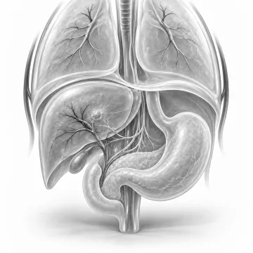
Palliative cancer care
Small procedures that ease fluid build-up, pain, blockages and eating problems caused by cancer.
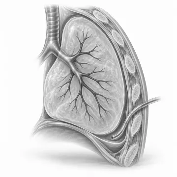
Pleurodesis
Lasting relief for fluid that keeps returning around the lung: talc pleurodesis or a home drain.
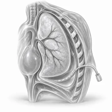
Chest PleurX catheter
A home drain for fluid that keeps building up around the lung.
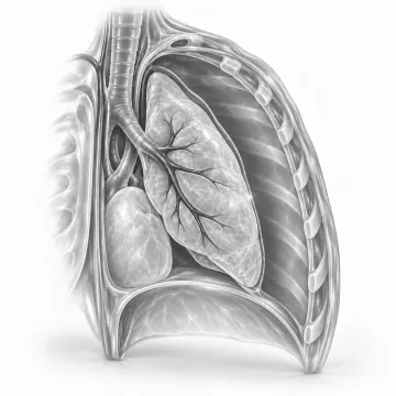
Pneumothorax (collapsed lung)
A collapsed lung: causes, symptoms, when it is an emergency and chest tube treatment.
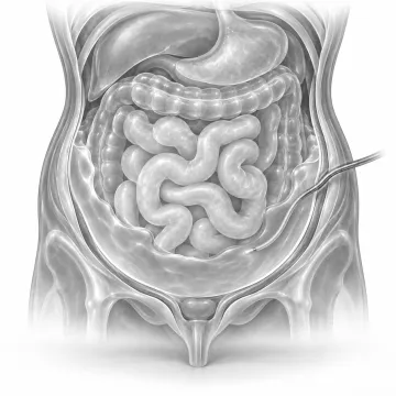
Paracentesis
Drain fluid from the belly through a thin tube, with numbing medicine and quick relief.
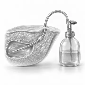
PleurX catheter
A tunneled home drain for fluid that keeps returning in the chest or belly.
Written for patients by the Palo Alto Interventional team. General information only; your own plan depends on your health and imaging.
Talk to us about your options
Request a consultation online, or call 650-404-8445, Monday to Friday, 8 am to 5 pm. You can also ask your doctor to send a referral. We will review your imaging and help you decide whether a procedure makes sense.