
What is interventional radiology?
Treatment through a pinhole, guided by imaging.
Interventional radiologists use live X-ray, ultrasound and CT to guide thin tubes and needles to the problem from the inside, usually through a cut smaller than a grain of rice. Most patients go home the same day.
- Pinhole access
- Live image guidance
- Often same-day
- Local or sedation
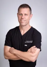 Fabio Komlos, MDInterventional radiologist
 Watch the video explanation3:26 video · Welcome to Vascular and Interventional Radiology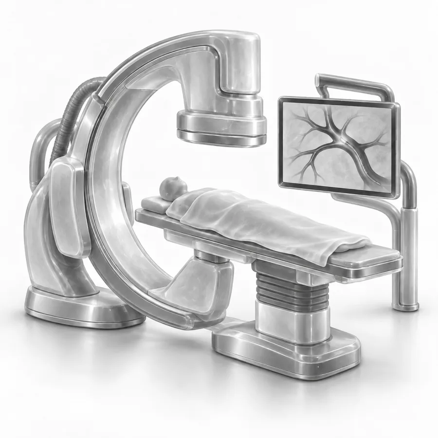
Live fluoroscopy from an IVC filter removal. We watch every move of the catheter on this screen.
Welcome to Vascular and Interventional Radiology
Dr. Fabio Komlos walks through what is interventional radiology? the way he explains it in clinic, using the same images he uses to plan treatment.
Fabio Komlos, MDInterventional radiologist · 3:26Everything you need to know about angiograms and what to expect after
13:56
Before your procedure
3:29
Quick answers
Under a minute eachWhat interventional radiology is
Interventional radiology (IR) is a medical specialty that treats disease from the inside of the body. Instead of opening the skin to see an organ, we use medical imaging to see it, and we reach it with thin, flexible tubes called catheters, fine wires and needles.
Most IR procedures start with a small puncture in the skin, often at the wrist, the upper thigh or directly over the target. Blood vessels act like a road map that leads to almost any organ. That is how we can stop bleeding, open blocked vessels, shrink tumors, drain infections or take a tissue sample without a surgical incision.
IR touches almost every part of medicine: blood vessels and veins, cancer care, the kidneys and bile ducts, pain and the spine, women's and men's health, and long-term access lines such as ports and PICC lines.
How image guidance works
Imaging is how we see what we are doing, in real time. We choose the tool that shows the target best and uses the least radiation.
- X-ray (fluoroscopy): a live X-ray video. With contrast, a dye that shows up on X-ray, the blood vessels light up like a map. This picture of the vessels is called an angiogram.
- Ultrasound: sound waves, no radiation. Ideal for finding a vein, guiding a needle into the thyroid or liver, or checking a fluid pocket.
- CT (computed tomography): cross-section X-ray images that show deep organs such as the lung, kidney or spine. We use it to place a needle within millimeters of a target.
- Cone-beam CT: the X-ray arm spins around you in a few seconds and builds a 3D picture during the procedure, so we can confirm exactly which vessels feed a tumor or organ.
We keep radiation as low as possible. We use short pulses of X-ray, tight framing on the area we need and ultrasound whenever it does the job. For most procedures the dose is similar to or lower than a few diagnostic CT scans.
Pinhole or surgery: how they compare
IR is not always better than surgery. For some problems surgery is the right answer, and for many there is a choice. Here is how the two approaches usually differ.
| IR procedure | Open surgery | |
|---|---|---|
| Opening in the skin | A 2–3 mm puncture, usually no stitches | An incision of several centimeters or more |
| Anesthesia | Local numbing, often with sedation | Usually general anesthesia |
| Hospital stay | Often home the same day | Often one or more nights |
| Back to normal | Usually days | Often weeks |
| Scar | Tiny or none | Visible scar |
Recovery depends on the procedure and on your overall health. Your doctor will explain what to expect for your specific treatment.
Who interventional radiologists are
Interventional radiologists are medical doctors (MD or DO). After 4 years of medical school, they complete about 6 to 7 more years of training: a year of general clinical medicine, then residency in diagnostic radiology and interventional radiology. Many also complete an extra fellowship year.
That training combines two skills that rarely live in one person: reading medical images like a radiologist, and treating patients with catheters and needles like a proceduralist. In the United States, interventional radiologists are certified by the American Board of Radiology.
Because we often work behind the scenes, many patients have never heard of us. You may meet us when your surgeon, oncologist, primary care doctor or the emergency team asks for help, or you can come to us directly for a consultation.
What the doctor sees and how we think through a case
Before your procedure, we study your scans in detail: where the problem is, which blood vessels supply it, what sits nearby and what the safest path looks like. We plan the entry point, the tools and a backup plan before you arrive.
During the procedure, the room has large screens showing live X-ray or ultrasound, your vital signs and your earlier scans side by side. We compare the live picture with the plan at every step. When we inject contrast, we watch how blood flows. When we place a needle, we confirm its tip is exactly where it should be before we do anything else.
When I look at a scan, I am asking three questions: what is the problem, what is the safest way to reach it, and what could surprise me on the way there. I treat the screen as a window into your body, and I do not move forward until what I see matches what I expect. I also like to show patients their own images afterward, because seeing it makes the whole thing much less mysterious.Fabio Komlos, MD, interventional radiologist · About Dr. Fabio Komlos
What to expect as a patient
Most IR procedures take 30 to 90 minutes. You are usually awake but relaxed, with the skin numbed so you feel pressure rather than pain. Learn more in our guides on sedation, getting ready and recovery at home.
If you take a blood thinner, tell us early, because it may need to be paused under a plan from us and your prescribing doctor. See blood thinners and procedures.
Common questions
Is an interventional radiologist a surgeon?
Will I be asleep?
Does it hurt?
How much radiation will I get?
Do I need a referral?
Will insurance cover an IR procedure?

Before your procedure
Fasting, medicines, blood thinners, diabetes, a driver and what to bring: your checklist.
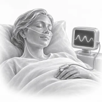
Sedation and anesthesia
Local numbing, twilight sedation and anesthesia: what you feel and why you need a ride.

After your procedure
Puncture site care, activity, showering, driving, pain and when to call after an IR procedure.
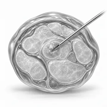
Image-guided biopsies
Get a tissue diagnosis through a needle, guided by ultrasound or CT, usually without an incision.
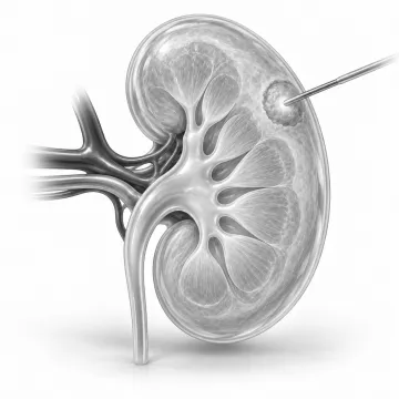
Tumor ablation
Heat or freeze a tumor through a needle, with no incision and a short recovery.
Written for patients by the Palo Alto Interventional team. General information only; your own plan depends on your health and imaging.
Talk to us about your options
Request a consultation online, or call 650-404-8445, Monday to Friday, 8 am to 5 pm. You can also ask your doctor to send a referral. We will review your imaging and help you decide whether a procedure makes sense.