
Pneumothorax (collapsed lung)
Help a collapsed lung re-expand, often with a small tube.
A pneumothorax is air leaking into the space around the lung, making it partly collapse. Small ones often heal on their own. Larger ones are treated with a thin chest tube, usually for 1 to 3 days.
- Sudden chest pain
- Common in young adults
- Chest tube 1–3 days
- Can come back
 Fabio Komlos, MDInterventional radiologist
 Watch the video explanation6:29 video · Lung Biopsy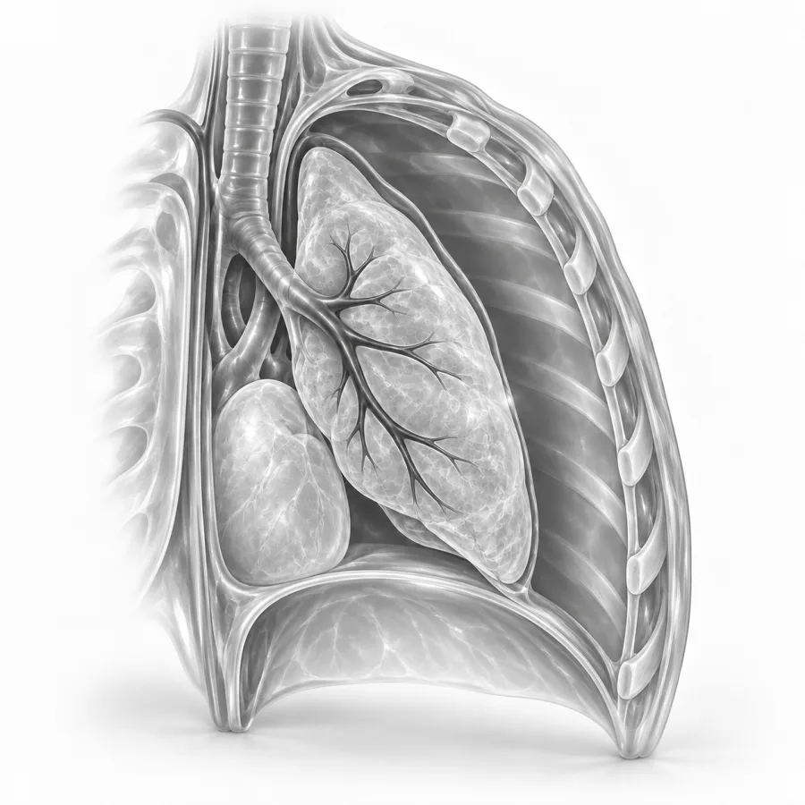
Breathing often improves within minutes of placement.
Lung Biopsy
Dr. Fabio Komlos walks through pneumothorax (collapsed lung) the way he explains it in clinic, using the same images he uses to plan treatment.
Fabio Komlos, MDInterventional radiologist · 6:29Thoracentesis Procedure: Everything You Need to Know
12:23
Lung Biopsy Prep: Five Critical Steps Before Your Procedure
3:16
What is a pneumothorax?
Your lungs are wrapped in a thin, two-layer lining called the pleura. Normally the two layers glide against each other with no air between them. A pneumothorax happens when air gets into that space, called the pleural space. The air pushes on the lung and makes part or all of it collapse, like a balloon that cannot fully inflate.
Why it happens
- Primary spontaneous: a small air blister on the lung surface, called a bleb, pops. This is most common in tall, thin young adults, especially men who smoke.
- Secondary spontaneous: an existing lung disease, such as COPD or emphysema, cystic fibrosis or a lung infection, weakens the lung.
- After a procedure: for example a lung biopsy, a central line or a thoracentesis. This is usually small.
- After an injury: a broken rib, a fall or a car accident.
On a chest X-ray, the edge of the collapsed lung shows up as a thin white line. Beyond it, the space is black with no lung markings, because it is filled with air.
Symptoms
- Sudden sharp or stabbing chest pain on one side, often worse with a deep breath
- Shortness of breath, from mild to severe
- A fast heartbeat or feeling anxious
- A dry cough
- Pain that spreads to the shoulder on the same side
A small pneumothorax can cause only mild pain and go unnoticed for a day or two. People with lung disease often feel much worse, even with a small one, because they have less lung reserve.
When it is an emergency
Rarely, air keeps entering the chest and cannot escape. Pressure builds up and pushes on the heart and the other lung. This is called a tension pneumothorax, and it is life-threatening. It needs immediate treatment to let the air out.
How it is diagnosed
A doctor listens to your chest; breath sounds may be quiet on the affected side. A chest X-ray usually confirms the diagnosis and shows how large the pneumothorax is. A CT scan gives more detail, finds small air pockets and can show blebs or lung disease. In emergency rooms, a quick ultrasound at the bedside can also detect it.
When I look at the chest X-ray, I measure how far the lung edge has pulled away from the chest wall and compare it with earlier images. Just as important is how you are breathing. A small pneumothorax in a comfortable young person may only need watching, while the same size in someone with emphysema may need a tube right away.Fabio Komlos, MD, interventional radiologist · About Dr. Fabio Komlos
Treatment options
| Observation | Needle aspiration | Small chest tube | Surgery (VATS) | |
|---|---|---|---|---|
| Best for | Small, few symptoms | Moderate, first episode | Larger, symptoms, or lung disease | Leak that will not stop, or repeat episodes |
| What it involves | Rest, oxygen, repeat X-rays | Air removed with a needle and syringe | Thin tube left in to let air out | Keyhole surgery to fix the leak and seal the lining |
| Hospital time | Often none or a few hours | Often home the same day | Home with a valve, or 1 to 3 days in hospital | Several days |
| Chance it comes back | Same as untreated | Same as untreated | Same as untreated | Much lower |
Interventional radiologists often place small-bore chest tubes, thin tubes about the width of a pencil lead to a drinking straw. They are guided by CT, ultrasound or X-ray and are more comfortable than the large tubes used in surgery. If the air leak lasts longer than about 4 to 5 days, or the lung collapses again, a chest surgeon may recommend video-assisted thoracoscopic surgery (VATS). The surgeon removes the blebs and makes the lining stick to the chest wall so it cannot collapse again. See also pleurodesis.
Chest tube placement, step by step
Chest tubes are placed at El Camino Hospital with moderate sedation and numbing medicine. Your heart rate, blood pressure and oxygen are watched throughout. The procedure usually takes 15 to 20 minutes.
- 1Getting ready
You lie on your back or side. Sedation through your IV helps you relax, and oxygen is given.
- 2Numbing and planning
We use imaging to choose a safe spot, usually high on the front of the chest or on the side. The skin and the space between the ribs are numbed.
- 3Placing the tube
A thin needle enters the pleural space, and a guidewire follows. A small, soft tube slides over the wire and curls inside the chest.
- 4Connecting and securing
The tube is stitched and taped to the skin, then connected to a one-way Heimlich valve or a drainage box with suction. Many people breathe easier right away.
Recovery and living with it
With a Heimlich valve, a small one-way valve on the end of the tube, many people can go home and return in a day or two for an X-ray. If the tube is connected to suction, you stay in the hospital. Chest X-rays show when the lung has re-expanded and the leak has stopped. Removal takes a few seconds: the stitch is cut, the tube slides out and an airtight dressing goes on for 24 hours.
- With the tubeSome soreness at the site. Walk around, breathe deeply and use pain medicine as needed. Keep the dressing dry.
- 1 to 3 daysMost air leaks stop. The tube is removed once X-rays show the lung is fully expanded.
- 2 to 4 weeksA follow-up X-ray is often done. Avoid heavy lifting, contact sports and strenuous exercise until you are cleared.
- FlyingWait until an X-ray shows the lung is fully re-expanded, and then usually at least 1 more week. Ask us before you book.
- Long termStop smoking; it greatly raises the chance of another collapse. Scuba diving is generally unsafe for life unless you have had surgery to prevent recurrence.
After a first spontaneous pneumothorax, about 1 in 3 people have another within a few years, most often in the first year. A pneumothorax after a lung biopsy or other procedure usually does not come back.
Common questions
Does a chest tube hurt?
Can I go home with a chest tube?
What if the tube comes out by accident?
When can I fly?
Will it happen again?
Is a pneumothorax after a lung biopsy serious?
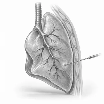
Lung biopsy
Find out what a lung nodule is with a CT-guided needle biopsy, usually home the same day.
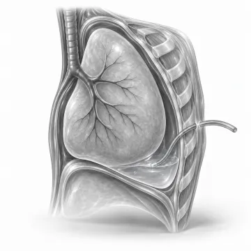
Thoracentesis
Drain fluid around the lung through a thin tube between the ribs, often with relief the same day.
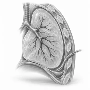
Pleurodesis
Lasting relief for fluid that keeps returning around the lung: talc pleurodesis or a home drain.
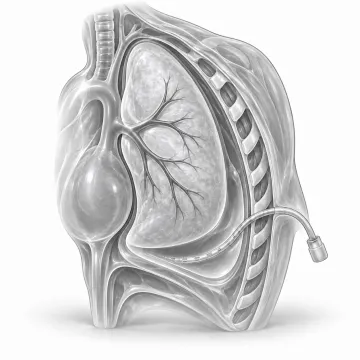
Chest PleurX catheter
A home drain for fluid that keeps building up around the lung.
Written for patients by the Palo Alto Interventional team. General information only; your own plan depends on your health and imaging.
Talk to us about your options
Request a consultation online, or call 650-404-8445, Monday to Friday, 8 am to 5 pm. You can also ask your doctor to send a referral. We will review your imaging and help you decide whether a procedure makes sense.