
Arteriovenous malformations (AVMs)
Understand and control an AVM, often without surgery.
An arteriovenous malformation (AVM) is a tangle of blood vessels where arteries connect straight to veins. Many can be controlled with embolization, a treatment done through a small tube in the wrist, groin or skin.
- Often present at birth
- Can grow over years
- MRI and ultrasound
- Staged treatment
 Fabio Komlos, MDInterventional radiologist
 Watch the video explanation3:47 video · What exactly is a vascular malformationand when should you worry?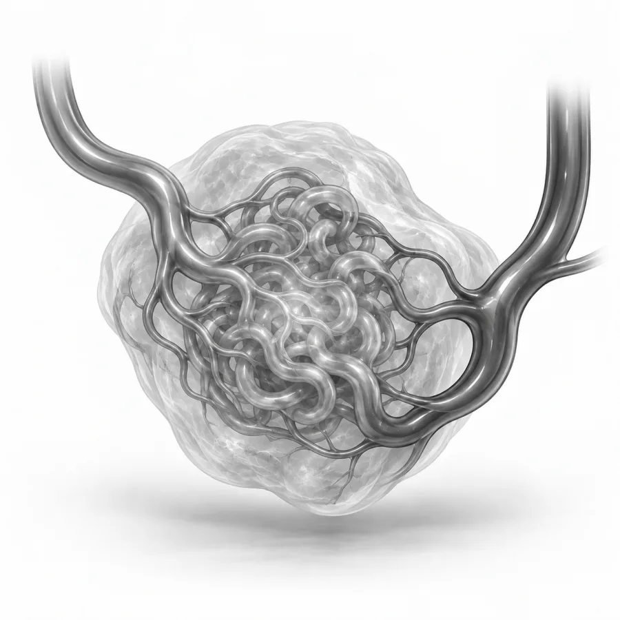
 On imaging
Angiogram during an embolization: the catheter tip sits in the target vessel before it is closed.
What exactly is a vascular malformationand when should you worry?
Dr. Fabio Komlos walks through arteriovenous malformations (avms) the way he explains it in clinic, using the same images he uses to plan treatment.
Fabio Komlos, MDInterventional radiologist · 3:47Vascular Malformations Explained — Types, Risks — Minimally Invasive Treatments
3:28
Quick answers
Under a minute eachWhat is an AVM?
Normally, blood flows from arteries into a fine mesh of tiny vessels called capillaries, and then into veins. The capillaries slow the blood down and feed the tissue. In an arteriovenous malformation (AVM), some arteries connect directly to veins, skipping the capillaries. High-pressure blood rushes straight into the veins, which stretch and enlarge over time.
The spot where arteries and veins meet is called the nidus (Latin for nest). A simpler AVM with a single direct connection is called an arteriovenous fistula. Most AVMs start before birth, even if they are not noticed until childhood, the teenage years or adulthood. They are not cancer and do not spread.
AVMs belong to a larger family of vascular malformations. Most others, such as venous or lymphatic malformations, have slow flow. AVMs are the fast-flow type, which is why they behave differently and need careful planning.
AVMs in the brain and spinal cord are managed by specialized neurovascular teams. This page focuses on AVMs in the rest of the body: arms, legs, pelvis, lungs, kidneys, face and other organs.
Symptoms
Symptoms depend on where the AVM is, how big it is and how much blood flows through it. Many start quietly and grow slowly over years. Growth often speeds up during puberty, pregnancy, after an injury or after incomplete surgery.
- A warm, pink or red patch of skin that may slowly swell
- A pulsing lump, or a buzzing feeling you can feel with your hand (called a thrill)
- Pain, heaviness or pressure in the area
- One arm or leg larger or longer than the other
- Skin sores that do not heal, or bleeding
- In the pelvis or kidney: blood in the urine or heavy bleeding
- In the lungs: shortness of breath, low oxygen, nosebleeds, or rarely a stroke or brain abscess
- In very large AVMs: heart strain, with breathlessness or leg swelling
Lung AVMs deserve a special mention. They let blood skip the lung's natural filter, so small clots or bacteria can pass to the brain. Many people with lung AVMs have an inherited condition called hereditary hemorrhagic telangiectasia (HHT), which also causes frequent nosebleeds and red spots on the lips and hands. If you have HHT, your family members should be screened too.
When to worry
Doctors often describe AVM stages to guide timing. A quiet AVM may be warm and pink but stable. An expanding AVM gets bigger and starts to pulse. A destructive AVM causes pain, sores or bleeding. A very large AVM can strain the heart. Treatment is usually considered once an AVM is growing or causing symptoms.
How we diagnose it
Our first goal is to confirm the diagnosis, because some tumors can look like vascular malformations. We then map the AVM in detail so we can plan the safest treatment.
- 1Ultrasound with Doppler. Painless and often done in the office. It shows fast flow and helps tell an AVM from a slow-flow malformation.
- 2MRI or CT scan. Shows how large the AVM is and which muscles, organs, nerves and bones it involves. For lung AVMs, a CT scan of the chest is the main test.
- 3Angiogram. A thin tube (catheter) is guided from the groin or wrist to the AVM. Contrast dye shows each feeding artery, the draining veins and how fast blood flows. This is often done as the first step of treatment.
What we seeTreatment options
Every AVM is different, so treatment is planned around its location, size, symptoms and your goals. For all but the simplest AVMs, your history, exam and imaging are reviewed by a team that includes interventional radiology and vascular surgery.
| Watchful waiting | Embolization | Surgery | |
|---|---|---|---|
| Best for | Small, quiet AVMs with no symptoms | Most growing or symptomatic AVMs, and lung AVMs | Small AVMs that can be fully removed safely |
| How it works | Regular checks and compression garments | Material injected through a catheter or needle blocks the nidus | The abnormal tissue is cut out |
| Incision | None | None, a small puncture | Yes |
| Trade-offs | AVM may grow over time | Often needs several sessions; swelling after | Can damage nearby muscle, nerves or organs; often done after embolization |
Embolization means blocking the abnormal vessels from inside. We guide a catheter through an artery, or place a needle directly into the AVM, and inject materials such as liquid glue-like agents, alcohol or tiny metal coils. The aim is to close the nidus itself, not just the arteries feeding it. Blocking only the feeding arteries tends to make the AVM recruit new ones and come back worse.
For lung AVMs, the feeding artery is usually closed with coils or plugs in a single session. Learn more on our AVM embolization page.
With an AVM, the most important thing I do happens before any treatment: I study the angiogram and MRI until I understand exactly where the nidus is and what surrounds it. I tell patients honestly that large AVMs are often controlled rather than cured, and that we will likely treat in stages. My goal is to make you feel better and keep the AVM from causing harm, while protecting the healthy tissue around it.Fabio Komlos, MD, interventional radiologist · About Dr. Fabio Komlos
What treatment is like
Embolization is usually an outpatient procedure, meaning you go home the same day. We do these procedures at El Camino Hospital in Mountain View, in a specialized procedure room with X-ray, ultrasound and CT imaging. Most are done with local numbing and moderate sedation, which makes you relaxed and drowsy. Some complex AVMs need general anesthesia.
A small tube is placed in the groin, wrist or directly into the malformation. There is no incision and no stitches. Afterward you are watched for at least 2 hours to make sure there is no bleeding before you go home.
- First few daysPain and swelling in the treated area are expected and can be significant. Anti-inflammatory medicine, pain medicine and sometimes a short course of steroids help.
- 1–2 weeksSwelling settles. Keep the area raised if it is an arm or leg. Watch the skin over the AVM.
- Monthly during treatmentWe usually see you in clinic about once a month while treatment is under way, and plan the next stage if needed.
- Long termPeriodic imaging to watch for regrowth. AVMs can recur, and a repeat treatment is sometimes needed.
Possible risks
- Pain and swelling, which are common
- Skin blistering, darkening or a sore over the treated area
- Numbness or weakness if a nearby nerve is affected, usually temporary
- Embolic material reaching a normal vessel and reducing blood flow to healthy tissue, which is rare
- Bleeding or bruising where the catheter entered
Living with an AVM
Outcomes vary a lot because AVMs vary a lot. Small AVMs, especially those with a single draining vein, can often be cured with embolization alone. Large, spread-out AVMs are usually not curable, but most people get meaningful relief of pain, bleeding and swelling with treatment.
- Wear compression garments on an arm or leg AVM if we recommend them.
- Protect the skin over the AVM from injury.
- Tell us before a planned pregnancy, since hormones can make AVMs grow.
- If you have HHT or a lung AVM, ask about screening for family members and about taking antibiotics before dental work.
Common questions
Can an AVM be cured?
Is an AVM cancer?
How many treatments will I need?
Will it hurt afterward?
Why not just remove it surgically?
I have a lung AVM but feel fine. Should it be treated?
Will insurance cover treatment?
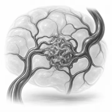
Arteriovenous malformation embolization
Close the abnormal artery-to-vein connections of an AVM through a pinhole, without surgery.
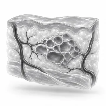
Vascular malformations
Venous, lymphatic and arteriovenous malformations: what they are and how they are treated.
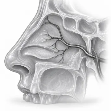
Epistaxis embolization
Stop severe or recurring nosebleeds by sealing the bleeding arteries from the inside.
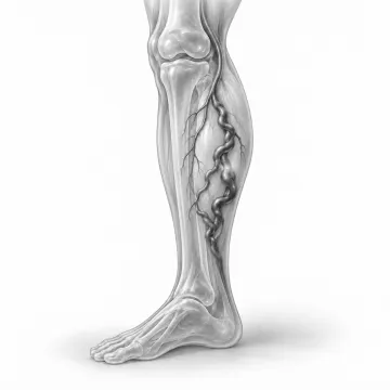
Varicose veins
Why leg veins bulge and ache, and how to treat them without major surgery.
Written for patients by the Palo Alto Interventional team. General information only; your own plan depends on your health and imaging.
Talk to us about your options
Request a consultation online, or call 650-404-8445, Monday to Friday, 8 am to 5 pm. You can also ask your doctor to send a referral. We will review your imaging and help you decide whether a procedure makes sense.