
Arteriovenous malformation embolization
Calm a problem AVM without open surgery.
Embolization closes the abnormal connections inside an arteriovenous malformation (AVM) from the inside, through a pinhole in the groin, wrist or skin. There are no incisions or stitches, and most people go home the same day.
- Outpatient
- 2–4 hours
- Sedation or anesthesia
- Often done in stages
 Fabio Komlos, MDInterventional radiologist
 Watch the video explanation3:47 video · What exactly is a vascular malformationand when should you worry?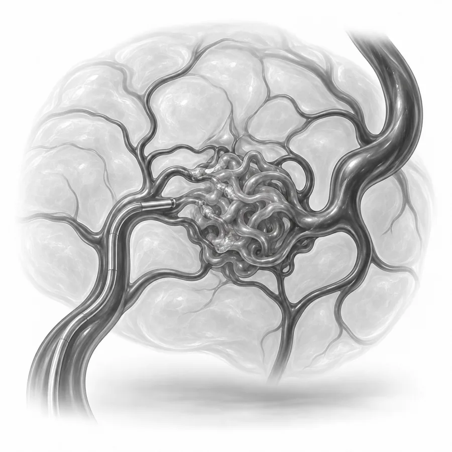
 On imaging
Angiogram during an embolization: the catheter tip sits in the target vessel before it is closed.
What exactly is a vascular malformationand when should you worry?
Dr. Fabio Komlos walks through arteriovenous malformation embolization the way he explains it in clinic, using the same images he uses to plan treatment.
Fabio Komlos, MDInterventional radiologist · 3:47Vascular Malformations Explained — Types, Risks — Minimally Invasive Treatments
3:28
After Embolization? Don't Make These 5 Mistakes
5:32
Everything you need to know about angiograms and what to expect after
13:56
Quick answers
Under a minute eachWhat problem does it treat?
Normally, blood flows from arteries into a web of tiny vessels called capillaries, which slow it down and feed the tissue, and then into veins. In an arteriovenous malformation (AVM), some arteries connect straight to veins, skipping the capillaries. The center of these abnormal connections is called the nidus, from the Latin word for nest. Blood rushes through at high pressure, and over time the veins stretch and the tissue around them can swell, hurt, break down or bleed.
AVMs are present from birth and can be found almost anywhere: the arms and legs, pelvis, face, lungs, kidneys or liver. This page covers AVMs outside the brain and spine, which are treated by interventional radiologists. Brain AVMs are usually treated by neurointerventional and neurosurgery teams. For the bigger picture, see arteriovenous malformations and vascular malformations.
Embolization means blocking blood vessels on purpose. For an AVM, the goal is to fill and seal the nidus and the start of the draining vein, so blood has to take the normal route again.
Who it helps and who it may not
Good candidates
- AVM causing pain, swelling or skin sores
- Bleeding from the skin, mouth, uterus or an organ
- A visible, warm, pulsing mass that is growing
- Heart strain from very high blood flow (rare)
- Lung AVMs, which can let clots reach the brain
- Before surgery, to reduce bleeding during removal
May not be right if
- The AVM is small and causes no symptoms
- The feeding arteries also supply vital normal tissue that cannot be protected
- Kidney problems or a serious allergy to contrast dye (can sometimes be managed)
- Pregnancy, in most cases, until after delivery
- Active infection in the area
Treating an AVM that causes no symptoms is not always wise. Partial treatment can sometimes make an AVM more active. That is why we weigh the risks carefully and treat when the benefit is clear, with one important exception: lung AVMs are often treated even without symptoms, because they carry a risk of stroke.
How it compares with the alternatives
| Embolization | Surgery | Watching | |
|---|---|---|---|
| How it's done | Catheter or needle, no incision | Cut out the malformation | Regular checks, compression |
| Anesthesia | Sedation or general | General | None |
| Recovery | Days to 2 weeks | Weeks, depending on area | None |
| Best for | Most symptomatic AVMs, especially deep or near nerves | Small AVMs that can be removed completely | Small AVMs with no symptoms |
| Downside | Often needs several sessions; can recur | Scars; risk to nearby structures; bleeding | AVM may slowly grow |
Surgery can cure an AVM if the whole nidus is removed, but when the AVM involves muscle, organs, nerves or tendons, surgery can cause more harm than good. Embolization and surgery are often combined, with embolization done 1 to 3 days before surgery to make the operation safer. Compression garments help with swelling and pain but do not treat the AVM itself.
Before the procedure
We start with an ultrasound, often in the office, and an MRI or CT scan to confirm the diagnosis and map the AVM. Complex cases are reviewed by a team that includes interventional radiology and vascular surgery. Sometimes we do a separate diagnostic angiogram first, to see exactly which arteries feed the AVM and how fast the flow is, and then bring you back for staged treatment.
How to prepare
- Do not eat for about 6 to 8 hours before the procedure. Small sips of water with your usual medicines are usually fine. We will give you exact instructions.
- Tell us about blood thinners, diabetes medicines, kidney problems and any allergy to contrast dye. See blood thinners.
- We may check blood work, including kidney function and clotting.
- Arrange for an adult to drive you home and, ideally, stay with you the first night.
- Read before your procedure and sedation.
What happens, step by step
- 1Getting comfortable
The procedure is done at El Camino Hospital in Mountain View, in a room with X-ray, ultrasound and CT imaging. You receive local numbing and twilight sedation, or general anesthesia for large, painful or complex AVMs.
- 2Mapping the AVM
A small tube is placed in the groin or wrist artery, or a thin needle is placed directly into the malformation. Contrast dye shows the feeding arteries, the nidus and the draining veins.
- 3Closing the connections
We deliver embolic agents into the nidus: liquid glues or gels that harden in place, alcohol that seals the vessel lining, or tiny coils and plugs. Sometimes a vein is temporarily blocked with a balloon to slow flow while we work.
- 4Checking and finishing
Repeat pictures confirm how much of the AVM is closed. The tubes are removed and pressure or a closure device seals the pinhole. No stitches are needed.
What we seeBefore every AVM embolization I study the angiogram frame by frame to see exactly where the arteries meet the veins, because that meeting point is what has to be closed. Blocking only the arteries feeding it is like closing a highway exit: the traffic just finds another route. I tell patients up front that we usually work in stages, and that my first priority is to protect the normal skin, nerves and muscle around the AVM.Fabio Komlos, MD, interventional radiologist · About Dr. Fabio Komlos
Recovery and aftercare
You are monitored for at least 2 hours to make sure there is no bleeding. Most people go home the same day. After a large treatment or general anesthesia, you may stay overnight.
- First 24 hoursRest with the treated area raised. No driving, alcohol or important decisions after sedation. You may shower the next day unless told otherwise.
- Days 2–5Pain and swelling are usually at their worst, and can be significant. Take prescribed pain medicine and anti-inflammatories, use ice, and keep the area raised. Sometimes steroids are given to calm the swelling.
- 1–2 weeksSwelling goes down and most people are back to normal activity. The area may feel firm as the treated vessels scar down. Avoid heavy lifting with a treated arm or leg until comfortable.
- About 1 monthClinic visit, and imaging if needed, to decide whether another session will help. Visits continue until symptoms settle.
Call us at 650-404-8445 if you have
- Fever above 101°F or chills
- Pain that keeps getting worse after day 3, or is not controlled by your medicines
- Skin over the treated area that turns dark, blisters or opens
- New numbness, weakness or coldness beyond the treated area
- Bleeding or growing swelling at the puncture site. For heavy bleeding, press firmly and call 911.
Results
Results depend a lot on the type of AVM. Small AVMs, especially those that drain into a single vein, have a good chance of being cured with embolization alone. Large, widespread AVMs are often not curable, and the goal is to control pain, bleeding and swelling. Most people have at least some improvement in their symptoms after treatment.
AVMs can come back, sometimes years later, as new vessels grow in. Repeat treatment is common and is part of the plan, not a failure. Hormone changes, injury and surgery can make an AVM more active, so long-term follow-up matters.
Risks
- Pain and swelling: expected, sometimes marked, usually improving within 1 to 2 weeks
- Skin injury: blistering, darkening or a skin sore over a treated AVM near the surface (uncommon)
- Nerve injury: numbness, tingling or weakness, usually temporary but occasionally lasting
- Non-target embolization: embolic material reaching normal vessels and harming the tissue they feed, or, rarely, traveling to the lungs (rare)
- Bleeding or bruising at the puncture site
- Contrast reactions or kidney strain
- Infection (rare)
Because every AVM is different, we will talk through the specific risks in your case before treatment.
Common questions
Does AVM embolization hurt?
How many sessions will I need?
Will my AVM be cured?
Will I have a scar?
When can I go back to work?
Can I have this if I am planning a pregnancy?
Will insurance cover it?
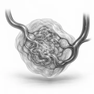
Arteriovenous malformations (AVMs)
What an AVM is, when it needs treatment, and how embolization can control it.
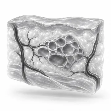
Vascular malformations
Venous, lymphatic and arteriovenous malformations: what they are and how they are treated.
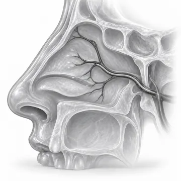
Epistaxis embolization
Stop severe or recurring nosebleeds by sealing the bleeding arteries from the inside.
Before your procedure
Fasting, medicines, blood thinners, diabetes, a driver and what to bring: your checklist.
Sedation and anesthesia
Local numbing, twilight sedation and anesthesia: what you feel and why you need a ride.
Written for patients by the Palo Alto Interventional team. General information only; your own plan depends on your health and imaging.
Talk to us about your options
Request a consultation online, or call 650-404-8445, Monday to Friday, 8 am to 5 pm. You can also ask your doctor to send a referral. We will review your imaging and help you decide whether a procedure makes sense.