
Vascular malformations
Shrink a vascular malformation without major surgery.
A vascular malformation is a tangle of blood or lymph vessels that formed the wrong way before birth. Many can be shrunk with needle injections or embolization, usually as a same-day procedure.
- Present from birth
- Can grow at any age
- Not cancer
- Treated in stages
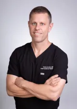 Fabio Komlos, MDInterventional radiologist
 Watch the video explanation3:28 video · Vascular Malformations Explained — Types, Risks — Minimally Invasive Treatments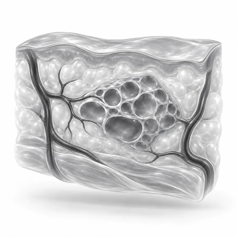
 On imaging
Angiogram during an embolization: the catheter tip sits in the target vessel before it is closed.
Vascular Malformations Explained — Types, Risks — Minimally Invasive Treatments
Dr. Fabio Komlos walks through vascular malformations the way he explains it in clinic, using the same images he uses to plan treatment.
Fabio Komlos, MDInterventional radiologist · 3:28What exactly is a vascular malformationand when should you worry?
3:47
Quick answers
Under a minute eachWhat is a vascular malformation?
Blood vessels and lymph vessels form in the early weeks of pregnancy. Sometimes a group of vessels forms the wrong way and ends up enlarged, tangled or connected in the wrong places. This is called a vascular malformation. It is present at birth, even if it is not noticed until childhood, the teen years or adulthood. It is not cancer and does not spread to other parts of the body.
Malformations grow along with the body and often enlarge during puberty, pregnancy, or after an injury or surgery. They do not go away on their own. They are different from a hemangioma, a common birthmark in babies that grows for several months and then usually shrinks by itself.
Most malformations happen by chance and are not inherited. A few are linked to gene changes, sometimes as part of a syndrome, and genetic testing can help guide treatment in those cases.
The main types
Doctors group malformations by which vessels are involved and how fast blood flows through them. The type decides the treatment.
| Venous | Lymphatic | Arteriovenous | |
|---|---|---|---|
| What it is | Enlarged, spongy veins | Fluid-filled pockets of lymph vessels | Arteries connected straight to veins, skipping the small vessels |
| Blood flow | Slow | No blood, clear fluid | Fast |
| How it looks | Soft, bluish, squishable; swells when hanging down | Soft lump, may be large; can swell with a cold | Warm, pink or red, often with a pulse or buzzing |
| Main treatment | Sclerotherapy | Sclerotherapy, sometimes medicine | Embolization, sometimes surgery |
- Venous malformations (VM) are the most common type. They often hurt after exercise or in the morning, because small clots form inside them.
- Lymphatic malformations (LM) can be made of large cysts (macrocystic), tiny ones (microcystic), or both. They may suddenly swell and hurt if they bleed inside or get infected.
- Arteriovenous malformations (AVM) are high-flow and tend to be the most active. See our page on arteriovenous malformations for more detail.
- Capillary malformations such as port-wine stains affect the skin only and are treated with laser by a dermatologist.
Symptoms
Symptoms depend on the type, size and location. A small malformation may cause no trouble at all. A larger one can affect how a limb works or how you feel about your appearance.
- A soft lump or swelling that may change size with position, exercise or the menstrual cycle
- Pain or aching, especially after activity
- Bluish, red or warm skin
- A limb that is larger or longer than the other
- Bleeding from the skin or, rarely, from inside organs
- Leg swelling or shortness of breath with a very large AVM, because the heart works harder (rare)
- Trouble breathing or swallowing, if the malformation is in the neck or mouth
When to worry
Pregnancy can make malformations bigger and, for venous malformations, raise the risk of blood clots. If you have a malformation and are planning a pregnancy, talk with us first.
How it's diagnosed
The first goal is to make sure it really is a vascular malformation and not a tumor, and to find out which type it is.
- Ultrasound, often done in the office, shows whether the lesion is solid or made of channels, and whether blood flows slowly or fast.
- MRI shows the full size of the malformation and which muscles, nerves and organs are involved. It is the key test for planning treatment.
- CT is sometimes used, especially for bone involvement or to map arteries.
- Angiogram: a thin tube is guided into the arteries and contrast dye is injected. It is the best way to see an AVM's feeding arteries and draining veins, and is usually done as part of treatment.
- Blood tests such as a D-dimer, a clot marker, can help in venous malformations.
What we seeTreatment options
Not every malformation needs treatment. We treat when it causes pain, bleeding, swelling that limits function, sores, or a change in appearance that bothers you. Treatment is planned for each person, and complex cases are reviewed by a team that includes interventional radiology and vascular surgery. Most treatments are done in stages, several weeks to months apart.
- Sclerotherapy: using ultrasound and X-ray to guide a thin needle through the skin into the malformation, we inject a medicine called a sclerosant that irritates the lining of the vessels so they scar down and shrink. It is the main treatment for venous and lymphatic malformations.
- Embolization: a thin catheter is guided through the arteries, or a needle is placed directly, to block the abnormal connections with glue-like liquids, plugs or coils. It is the main treatment for AVMs. Read more on our arteriovenous malformation embolization page.
- Surgery: best for small malformations that can be removed completely without harming nearby muscles, nerves or tendons. It is sometimes done after embolization to reduce bleeding.
- Laser or cryoablation: used for some small or skin-level malformations.
- Compression garments and medicines: compression sleeves or stockings ease swelling and pain in limb venous malformations. Low-dose aspirin or a blood thinner may reduce painful clots. A medicine called sirolimus can shrink some complex lymphatic and venous malformations.
When I see someone with a vascular malformation, I start by listening to what bothers them most, because the goal is to fix that, not to erase every vessel on the MRI. I look at the images to see how fast the blood flows and how close the malformation is to nerves, skin and muscle. I am honest that many malformations are controlled rather than cured, and that we usually work in stages to get the most benefit with the least risk.Fabio Komlos, MD, interventional radiologist · About Dr. Fabio Komlos
What sclerotherapy is like
- 1Preparation
The procedure is done as an outpatient at El Camino Hospital, with numbing medicine and moderate sedation, or general anesthesia for large or sensitive areas and for children.
- 2Finding the channels
Using ultrasound, we place a thin needle into the malformation and inject contrast dye to see how it drains.
- 3Treating
The sclerosant, sometimes mixed with air into a foam, is injected slowly while we watch on X-ray. Several spots may be treated in one session.
- 4Going home
The needle is removed and a bandage or compression wrap is applied. After about 2 hours of monitoring, most people go home the same day.
- First 2–3 daysSwelling and soreness peak. It can be significant. Rest, elevate the area, use ice and take the pain medicine prescribed. Sometimes a short course of steroids helps.
- First 1–2 weeksSwelling settles. The area may feel firm or lumpy as the vessels scar down. Return to normal activity as comfort allows.
- 1–3 monthsThe treated area keeps shrinking. We usually see you in clinic and decide whether another session is needed.
Results, risks and living with it
Most people have less pain and swelling after treatment, often after 1 to 3 sessions. Small, well-defined malformations can sometimes be cured. Large or widespread ones are usually controlled rather than cured, and they can regrow over years, so repeat treatment is common.
Possible risks
- Pain and swelling, which are expected and may last a week or more
- Blistering, skin darkening or a skin sore over the treated area
- Nerve irritation causing numbness or weakness, usually temporary
- Treatment material reaching nearby normal vessels (non-target embolization), which is rare
- Blood clots in deep veins, or kidney strain from breakdown of blood cells with some sclerosants
- Infection, which is rare
Living with a malformation often means long-term follow-up. Wear compression garments if we recommend them, stay active, and let us know about changes in size, pain or skin color. Tell any doctor treating you, including your dentist or obstetrician, that you have a vascular malformation.
Common questions
Is a vascular malformation cancer?
Will it go away on its own?
How many treatments will I need?
Does sclerotherapy hurt?
Can children be treated?
Will insurance cover treatment?
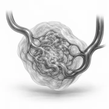
Arteriovenous malformations (AVMs)
What an AVM is, when it needs treatment, and how embolization can control it.
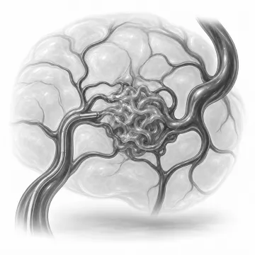
Arteriovenous malformation embolization
Close the abnormal artery-to-vein connections of an AVM through a pinhole, without surgery.
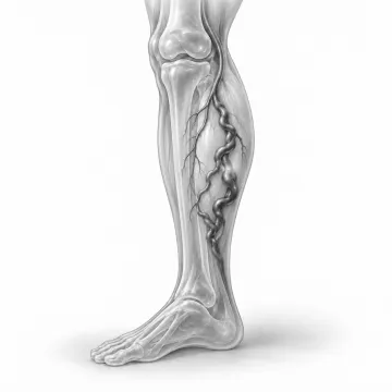
Varicose veins
Why leg veins bulge and ache, and how to treat them without major surgery.
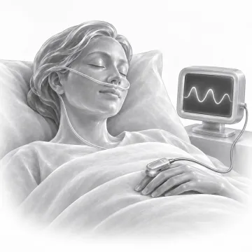
Sedation and anesthesia
Local numbing, twilight sedation and anesthesia: what you feel and why you need a ride.
Written for patients by the Palo Alto Interventional team. General information only; your own plan depends on your health and imaging.
Talk to us about your options
Request a consultation online, or call 650-404-8445, Monday to Friday, 8 am to 5 pm. You can also ask your doctor to send a referral. We will review your imaging and help you decide whether a procedure makes sense.