
Salivary gland biopsy
Know what the lump near your jaw is.
An ultrasound-guided salivary gland biopsy takes a small sample of a lump in the parotid gland, in front of the ear, or the submandibular gland, under the jaw. It takes about 20 to 30 minutes with numbing medicine.
- Outpatient
- 20–30 minutes
- Numbing medicine
- Drive yourself home
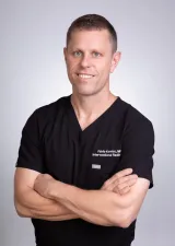 Fabio Komlos, MDInterventional radiologist
 Watch the video explanation12:30 video · How do you know if you have cancer or not?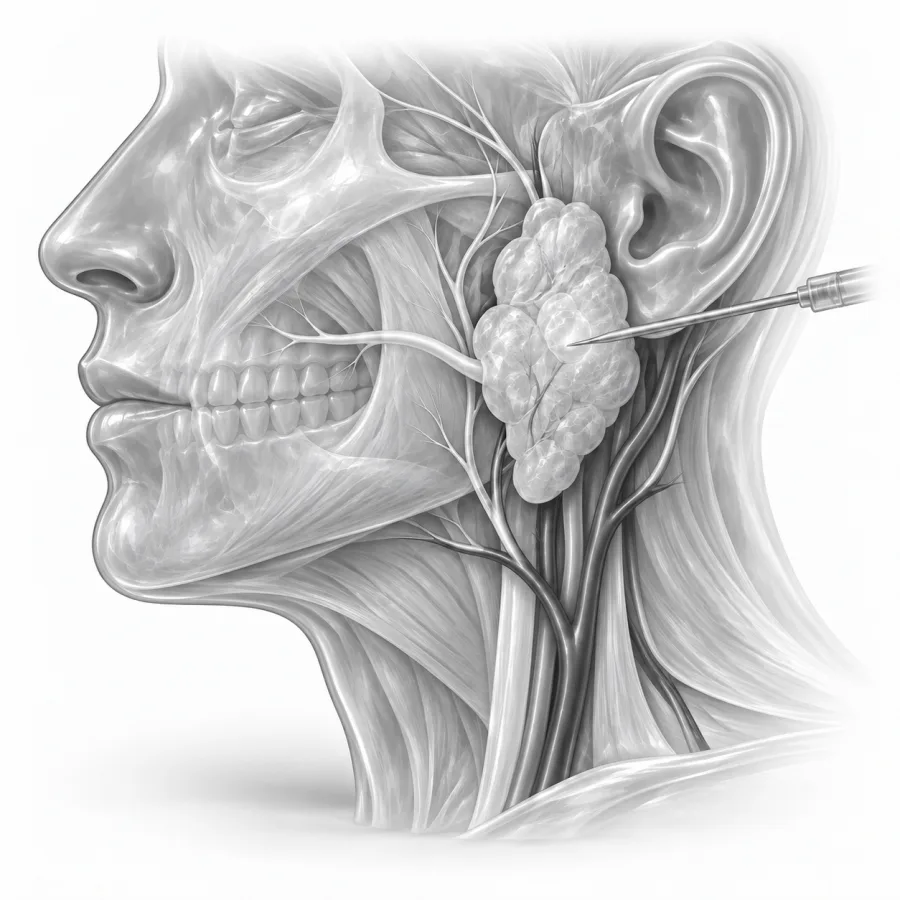
A thin needle guided by live ultrasound.
How do you know if you have cancer or not?
Dr. Fabio Komlos walks through salivary gland biopsy the way he explains it in clinic, using the same images he uses to plan treatment.
Fabio Komlos, MDInterventional radiologist · 12:30Swollen Lymph Node? When a Biopsy Is Necessary
10:56
Salivary glands and salivary gland lumps
You have three pairs of major salivary glands. The parotid glands sit in front of each ear and over the angle of the jaw. The submandibular glands sit under the jawbone. The sublingual glands lie under the tongue. Hundreds of tiny minor salivary glands line the mouth and throat.
A lump in one of these glands is often noticed while shaving or washing, or found on a scan. Many are harmless. About 4 out of 5 parotid tumors are benign, most commonly a pleomorphic adenoma or a Warthin tumor. Lumps in the submandibular gland are less common but more often cancer, close to half. Other causes include swollen lymph nodes inside the gland, cysts, stones and inflammation.
On ultrasound, the parotid gland looks like a bright, fine-textured gray area just under the skin in front of the ear. A tumor is usually a darker, rounded spot within it. We can see blood vessels around it and plan a path that avoids them.
Why a biopsy helps
Most salivary gland tumors, even benign ones, are eventually removed with surgery because they tend to keep growing. So why biopsy first? Because the type of tumor changes the plan. A biopsy helps your surgeon decide how much of the gland to remove, whether lymph nodes in the neck also need attention, and whether surgery is needed at all.
- Some lumps, such as a swollen lymph node from infection or a lymphoma, are treated without gland surgery.
- Warthin tumors in older adults can sometimes be watched instead of removed.
- If cancer is found, the surgeon can plan a larger operation from the start, and an oncologist can join the team early.
- In people who are not good candidates for surgery, a diagnosis still guides other treatment.
The biopsy is usually requested by an ear, nose and throat (ENT) surgeon after a CT, MRI or ultrasound.
Core needle or fine needle
| Fine needle aspiration (FNA) | Core needle biopsy | |
|---|---|---|
| Needle | Very thin, draws out cells | Slightly thicker, takes a thin strip of tissue |
| Accuracy | Good, but cells alone can be hard to classify | Higher; shows how the tissue is built |
| Best for | Small or cystic lumps, first test | Solid lumps, suspected lymphoma or cancer |
| Discomfort | Minimal | Mild, with good numbing |
We often use both in one visit. Salivary gland results are reported using a system called the Milan system, which sorts results into groups from nondiagnostic to malignant, much like thyroid results.
Salivary gland tumors come in dozens of types, and a small cluster of cells does not always tell them apart. That is why I often take a thin core as well as fine needle samples when the lump is solid. I map the blood vessels on ultrasound first and keep the needle in the outer part of the gland, away from where the facial nerve runs.Fabio Komlos, MD, interventional radiologist · About Dr. Fabio Komlos
How to prepare
- 1Tell us about any blood thinners. Many people can keep taking them for this biopsy, but we will confirm. See blood thinners.
- 2You can eat and drink normally unless you are having sedation.
- 3Bring any recent CT, MRI or ultrasound reports from other hospitals.
- 4Men may want to shave the area the morning of the biopsy.
What happens during the biopsy
- 1Positioning
You lie on your back with your head turned to the side away from the lump.
- 2Ultrasound and numbing
We find the lump with ultrasound, clean the skin and numb it and the tissue around the lump.
- 3Taking the samples
Watching on ultrasound, we take a few fine needle samples, a thin core, or both. You feel pressure, and a core needle makes a click.
- 4Finishing up
We press on the site for several minutes to limit bruising and apply a small bandage or skin glue.
Recovery, results and risks
- Right afterRest for about 30 minutes with an ice pack. You can eat, drink and drive yourself home if you had no sedation.
- First 1–2 daysMild soreness or swelling, sometimes worse with chewing or eating sour foods, which make the gland work. You can shower the same day.
- Up to 2 weeksAny bruise fades. Skin glue falls off on its own.
Results usually take about 2 to 5 business days and go to the surgeon or doctor who ordered the biopsy. Some tumors need extra stains or genetic tests, which can take longer.
This biopsy is very safe. A bruise or small swelling is the most common side effect. Serious bleeding and infection are rare. Injury to the facial nerve, which moves the face, is extremely rare with an ultrasound-guided needle. Spread of tumor cells along the needle track has been a concern with some tumors, but it is very rare with modern thin needles. A sample that does not give an answer happens in a small share of cases.
Common questions
Does it hurt?
Could the needle damage my facial nerve?
If most tumors get removed anyway, why biopsy?
Is a Warthin tumor dangerous?
Can I eat normally afterward?
Will insurance cover it?
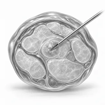
Image-guided biopsies
Get a tissue diagnosis through a needle, guided by ultrasound or CT, usually without an incision.
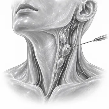
Lymph node biopsy
Diagnose a swollen lymph node with a needle guided by ultrasound or CT, usually without surgery.
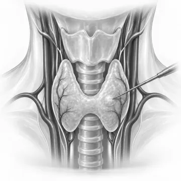
Thyroid biopsy
Check a thyroid nodule with a thin needle guided by ultrasound, usually with numbing medicine only.
Written for patients by the Palo Alto Interventional team. General information only; your own plan depends on your health and imaging.
Talk to us about your options
Request a consultation online, or call 650-404-8445, Monday to Friday, 8 am to 5 pm. You can also ask your doctor to send a referral. We will review your imaging and help you decide whether a procedure makes sense.