
Pulmonary embolism (blood clots in the lungs)
Clear dangerous lung clots and ease strain on the heart.
A pulmonary embolism (PE) is a blood clot that has traveled to the lungs, usually from a leg vein. Most are treated with blood thinners; for large clots, we can pull the clot out through a thin tube in a vein.
- Can be an emergency
- CT scan diagnosis
- Blood thinners for all
- Clot removal if large
 Fabio Komlos, MDInterventional radiologist
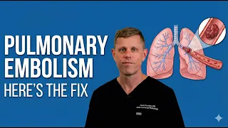 Watch the video explanation5:08 video · How Blood Clots Become Deadly Pulmonary Embolism — Modern Treatment Options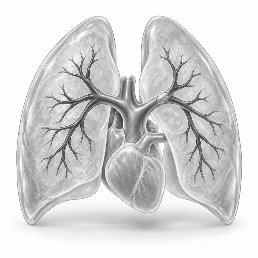
For sudden breathlessness, chest pain or fainting.
How Blood Clots Become Deadly Pulmonary Embolism — Modern Treatment Options
Dr. Fabio Komlos walks through pulmonary embolism (blood clots in the lungs) the way he explains it in clinic, using the same images he uses to plan treatment.
Fabio Komlos, MDInterventional radiologist · 5:08Pulmonary Embolism & Thrombectomy
12:19
Best Device to Treat Pulmonary Emboli in 2026
8:24
Quick answers
Under a minute eachWhat is a pulmonary embolism?
A pulmonary embolism (PE) happens when a blood clot blocks one or more arteries in the lungs. Almost always, the clot started in a deep vein of the leg or pelvis, a condition called deep vein thrombosis (DVT). A piece breaks off, travels through the heart and lodges in the lung arteries.
A clot in the lungs does two kinds of harm. It blocks part of the lung from picking up oxygen, so you feel short of breath. And it makes the right side of the heart, which pumps blood into the lungs, push against a blockage. A large clot can strain that side of the heart to the point of failure, which is why a big PE is an emergency.
The risk factors are the same as for DVT: recent surgery or hospital stay, not moving for long periods, cancer, pregnancy and the weeks after delivery, estrogen-containing hormones, a past clot and inherited clotting tendencies.
Symptoms and when it's an emergency
Common symptoms
- Shortness of breath that comes on suddenly or keeps getting worse
- Sharp chest pain that is worse when you breathe in or cough
- A fast heartbeat
- Coughing, sometimes with blood
- Lightheadedness or fainting
- Swelling or pain in one leg, from the DVT that started it
Some people have only mild breathlessness on stairs or feel unusually tired. Small clots can cause few symptoms.
How a PE is diagnosed
In the emergency room, the main test is a CT pulmonary angiogram: a CT scan of the chest taken while contrast dye flows through the lung arteries. It takes only a few minutes. Clots show up as dark gaps in the bright arteries. If you cannot have contrast dye, a lung scan called a V/Q scan may be used instead.
Your team then works out how serious the PE is. They check your blood pressure and oxygen, look at the size of the right side of the heart on the CT or an echocardiogram (heart ultrasound), and measure blood tests that show heart strain, such as troponin and BNP. A leg ultrasound may look for the DVT the clot came from.
On the CT scan, the lung arteries normally fill bright white with contrast. A clot appears as a dark gray plug inside the white. A large clot may sit where the main artery splits into left and right, called a saddle embolus.
| Low risk | Intermediate risk | High risk | |
|---|---|---|---|
| What it means | Normal blood pressure and no heart strain | Normal blood pressure but signs of heart strain | Low blood pressure or shock |
| Where treated | Sometimes at home or a short stay | Hospital, often a monitored unit | Intensive care |
| Main treatment | Blood thinners | Blood thinners, catheter treatment for some | Clot-dissolving medicine or clot removal right away |
Treatment options
Blood thinners (anticoagulants) are the foundation for everyone with a PE. They stop the clot from growing and stop new ones from forming while your body dissolves the clot over weeks to months. Treatment usually starts in the hospital with an injection or IV drip, then switches to pills such as apixaban (Eliquis) or rivaroxaban (Xarelto), or to warfarin. Most people take them for at least 3 months, and some longer. See our blood thinners guide.
For larger clots that strain the heart, doctors may also try to clear the clot faster. Many hospitals have a PE response team, where lung, heart, emergency and interventional specialists decide together on the best plan.
- Systemic thrombolysis: a strong clot-dissolving medicine given through an IV. It works fast but carries a real risk of serious bleeding, including in the brain, so it is kept mainly for people in shock.
- Catheter-directed thrombolysis: a much smaller dose of clot-dissolving medicine dripped directly into the lung arteries through thin tubes, usually over several hours.
- Catheter thrombectomy: the clot is pulled out through a tube with suction or a device, often with no clot-dissolving medicine at all.
- IVC filter: a small device that catches clots in the main abdominal vein, used mainly when you cannot take blood thinners. See IVC filters.
What catheter clot removal is like
Catheter treatment is done by an interventional radiologist in a procedure room with X-ray imaging. It is usually done with moderate sedation, which keeps you relaxed and drowsy, and numbing medicine at the skin. There is no incision.
- 1Access
We numb the skin at the groin or neck and place a thin tube into a vein using ultrasound.
- 2Measure and map
The tube is guided through the heart into the lung arteries. We measure pressures and inject contrast to see the clots.
- 3Remove the clot
Using suction catheters or a clot-removal device, we pull out as much clot as is safe. Sometimes we leave small tubes in place to drip clot-dissolving medicine for several hours instead.
- 4Finish and recover
The tubes are removed and the site is closed with a stitch or pressure. You recover in a monitored bed and continue blood thinners.
Many people feel their breathing ease within hours. Most go home after 1 to 3 days, depending on how sick they were before the procedure.
Possible risks
- Bleeding or bruising at the puncture site
- Bleeding elsewhere, higher if clot-dissolving medicine is used
- Irregular heartbeat while the catheter passes through the heart, usually brief
- Rarely, injury to a heart valve or lung artery
When I am called about a PE, the first thing I look at is not the clot, it is the right side of the heart. If the heart is coping, blood thinners usually do the job. If it is straining and the patient is getting worse, I talk with the team about going in to remove the clot. Watching someone's oxygen level climb and their heart rate settle on the table is one of the most satisfying parts of my work.Fabio Komlos, MD, interventional radiologist · About Dr. Fabio Komlos
Recovery and living after a PE
- First daysBreathlessness and chest discomfort start to ease. Walking is encouraged. You leave the hospital on a blood thinner.
- First weeksTiredness and breathlessness with effort are common. Increase activity gradually. Most people return to desk work within a few weeks.
- About 3 monthsFollow-up visit to decide how long to continue blood thinners. If you are still short of breath, you may have a heart ultrasound or lung scan.
- Long termMost people recover fully. A small number develop lasting breathlessness from scarred lung arteries, which can be treated.
A few people are left with long-term breathlessness because the clots do not fully dissolve and scar the lung arteries. This is called chronic thromboembolic pulmonary hypertension (CTEPH). It is uncommon, about 2 to 4 in 100 people after a PE, but it is important to catch because it can be treated with medicine, surgery or a balloon procedure.
- Take your blood thinner every day, exactly as prescribed.
- Keep moving. Short daily walks help your recovery and lower the chance of new clots.
- Tell your doctor if you are still short of breath 3 months after the PE.
- Plan ahead for long trips, surgery and pregnancy, since these raise clot risk.
Common questions
Is a pulmonary embolism always an emergency?
Will the clot go away?
Why remove a clot if blood thinners work?
How long will I be on blood thinners?
When can I exercise or fly?
Why am I still short of breath?
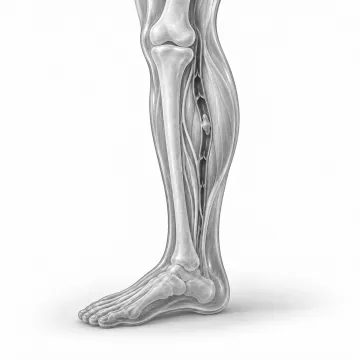
DVT (deep vein blood clots)
Blood clots in the leg or arm: signs, blood thinners, clot removal and long-term care.
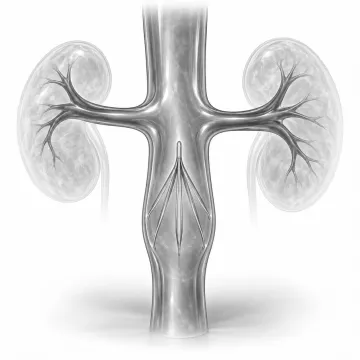
IVC filter placement and removal
Why IVC filters are placed, how they are removed, and why removal matters.
Blood thinners and your procedure
Which blood thinners may be paused before a procedure, typical hold times, and how bridging works.
Sedation and anesthesia
Local numbing, twilight sedation and anesthesia: what you feel and why you need a ride.
Written for patients by the Palo Alto Interventional team. General information only; your own plan depends on your health and imaging.
Talk to us about your options
Request a consultation online, or call 650-404-8445, Monday to Friday, 8 am to 5 pm. You can also ask your doctor to send a referral. We will review your imaging and help you decide whether a procedure makes sense.