
Endoleaks
Keep your aneurysm repair working for life.
An endoleak is blood still flowing into an aneurysm after a stent-graft repair (EVAR). Many are harmless and seal on their own. Those that let the aneurysm grow can usually be fixed through a pinhole.
- Found on routine scans
- Usually no symptoms
- Many seal on their own
- Pinhole treatment
 Fabio Komlos, MDInterventional radiologist
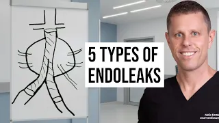 Watch the video explanation10:37 video · Endoleaks Explained: A Guide for Patients and Doctors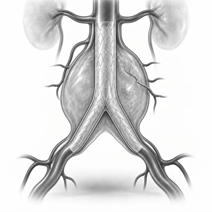
 On imaging
CT angiogram of the aorta and leg arteries, shown dark on light the way we read it.
Endoleaks Explained: A Guide for Patients and Doctors
Dr. Fabio Komlos walks through endoleaks the way he explains it in clinic, using the same images he uses to plan treatment.
Fabio Komlos, MDInterventional radiologist · 10:37Understanding Endoleaks After Aneurysm Repair — Dr. Fabio Komlos, Interventional Radiologist
5:35
What Is an Endoleak? A Common Cause of AAA Repair Failure
1:56
After Embolization? Don't Make These 5 Mistakes
5:32
What is an endoleak?
Endovascular aneurysm repair (EVAR) places a fabric-covered metal tube, called a stent-graft, inside an aortic aneurysm. Blood flows through the tube, and the old aneurysm around it, called the sac, is left without pressure. Over time the sac usually shrinks.
An endoleak means blood is still getting into the sac, outside the stent-graft. The word does not mean blood is leaking out of your body or into your belly. It is a leak into the space that the repair was meant to seal off. If enough pressure builds up in the sac, the aneurysm can keep growing and, rarely, burst. That is why every EVAR is followed by lifelong scans.
Endoleaks are common. Roughly 1 in 4 people who have EVAR will have one at some point, most often in the first year. Many need no treatment at all.
The five types
Doctors sort endoleaks into five types based on where the blood is coming from. The type tells us how urgent it is.
- Type I: blood slips around the top (Ia) or bottom (Ib) end of the stent-graft because the seal against the artery wall is not tight. The sac sees high pressure, so type I is treated promptly.
- Type II: the most common type. Small side branches of the aorta, such as lumbar arteries in the back or the inferior mesenteric artery that feeds the colon, keep filling the sac from behind. About half seal on their own. Treatment is usually needed only if the sac keeps growing.
- Type III: blood leaks through a hole in the fabric or where two pieces of the stent-graft join and have pulled apart. Like type I, this is high pressure and is treated promptly.
- Type IV: blood seeps through the fabric pores right after the procedure while you are on blood thinners. It usually stops on its own within about a month.
- Type V (endotension): the sac keeps growing but no leak can be seen on any scan. It is uncommon and is often re-studied with a different type of scan to look for a hidden leak.
What we seeSymptoms and when to worry
Most endoleaks cause no symptoms. They are found on the routine scans after EVAR, which is why keeping those appointments matters so much. You cannot feel whether your repair is working.
How endoleaks are found and followed
After EVAR you will have a scan at about 1 month, then usually at 12 months, and then every year if everything looks stable. The scan compares the size of the sac with earlier scans and looks for contrast outside the stent-graft.
- CT angiography (CTA): the most detailed picture, with contrast dye and several phases of images to catch slow leaks. It uses radiation and contrast, so it is not always the choice for every yearly check.
- Duplex ultrasound: no radiation and no dye. It measures the sac and can see many leaks, and it is often used for yearly follow-up once things are stable. Ultrasound with a special contrast (microbubbles) can find leaks that CT misses.
- MRI or catheter angiogram: used when the other tests do not explain why the sac is growing, or to plan treatment.
The number that matters most is the sac size. A sac that is shrinking or stable is reassuring, even if a small type II leak is present. A sac that has grown by 5 mm or more usually means it is time to treat.
Treatment options
The goal is to stop blood from pressurizing the sac. The right treatment depends on the type of leak.
| Type I or III | Type II | |
|---|---|---|
| Urgency | Treat promptly | Watch; treat if sac grows |
| Usual fix | Extra stent-graft pieces, balloon molding, or anchors to improve the seal | Embolization of the feeding branches or the sac |
| Approach | Through the groin arteries | Through the groin, or with a needle through the back |
| If that fails | Open surgery to repair or remove the graft | Repeat embolization, laparoscopic clipping, or surgery |
Embolization is the most common treatment for a type II endoleak. Embolization means blocking a blood vessel on purpose. We fill the leaking space, and the small arteries feeding it, with tiny soft metal coils and a liquid plug or glue. There are two main ways to reach it:
- Transarterial: a thin catheter passes from the groin through small arteries that connect to the leak, like driving through back roads to reach the source.
- Translumbar: you lie face down, and using CT or X-ray guidance we pass a thin needle through the skin of your back directly into the sac. This is often the most direct route.
When I read a scan after EVAR, the first thing I compare is the sac size against every earlier scan. A small type II leak with a sac that is shrinking does not worry me, and I tell patients that plainly. If the sac is growing, I study the delayed images carefully to find exactly which small arteries are feeding it and plan the most direct route in, because the aim is to seal the whole channel, not just one branch.Fabio Komlos, MD, interventional radiologist · About Dr. Fabio Komlos
What embolization is like
- 1Preparation
You do not eat for about 6 hours beforehand, and we give you instructions about blood thinners. An IV is placed and you receive moderate sedation or, sometimes, general anesthesia.
- 2Reaching the leak
We numb the skin and reach the sac either through a pinhole in the groin artery or with a thin needle through the back, guided by X-ray or CT.
- 3Sealing the leak
We place coils and a liquid embolic agent into the sac and the feeding branches until contrast no longer flows in. This usually takes 1 to 3 hours.
- 4Recovery
The catheter or needle is removed and the site is bandaged. You rest for a few hours, and some people stay overnight for observation.
- First dayRest at home with an adult to drive you. No driving, alcohol or important decisions for 24 hours after sedation. Mild back or groin soreness is common.
- First few daysSome people have a low fever or aching for a few days as the sac clots. Over-the-counter pain relievers usually help. Avoid heavy lifting for about a week.
- About 1 monthA follow-up scan checks that the leak has sealed and the sac is stable.
- Every yearLifelong scans continue, because a new leak can appear later.
Results and risks
Embolization stops sac growth in many patients, but type II endoleaks can come back through other small branches, and some people need a second treatment. Type I and III leaks are usually fixed well with extra stent-graft pieces. If less invasive fixes do not work, open surgery is an option, done by a vascular surgeon.
Possible risks
- Bruising or bleeding at the groin or back puncture
- Embolic material going somewhere it was not meant to go, such as the colon or spinal cord arteries (rare)
- Pain, low fever and tiredness for a few days
- Kidney strain from contrast dye
- Infection of the graft, which is rare but serious
Call us if you notice
- Fever above 101°F that lasts more than 2 days, or shaking chills
- Increasing belly or back pain
- Blood in the stool, or leg weakness or numbness
- Growing swelling or bleeding at the puncture site
Common questions
Is an endoleak dangerous?
Did my repair fail?
Will it seal on its own?
Does the procedure hurt?
Why do I need scans for the rest of my life?
Can I stop having CT scans because of the radiation?
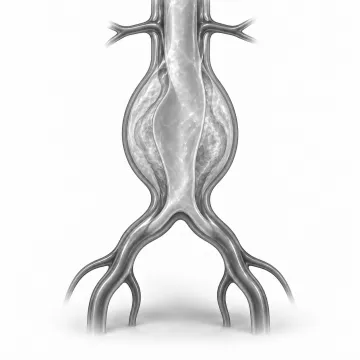
Aneurysms
A bulging artery found on a scan? What it means, when to watch and when to repair.
Peripheral arterial disease
Leg pain when walking or wounds that won't heal? Learn how blocked leg arteries are treated.

Before your procedure
Fasting, medicines, blood thinners, diabetes, a driver and what to bring: your checklist.

After your procedure
Puncture site care, activity, showering, driving, pain and when to call after an IR procedure.
Written for patients by the Palo Alto Interventional team. General information only; your own plan depends on your health and imaging.
Talk to us about your options
Request a consultation online, or call 650-404-8445, Monday to Friday, 8 am to 5 pm. You can also ask your doctor to send a referral. We will review your imaging and help you decide whether a procedure makes sense.