
Middle meningeal artery embolization
Help a chronic brain bleed heal and stay away.
Middle meningeal artery (MMA) embolization treats a chronic subdural hematoma by closing the fragile vessels that keep it bleeding, through a pinhole in the wrist or groin. It lowers the chance of the blood collection coming back.
- Hospital procedure
- 1–2 hours
- Sedation or anesthesia
- Often 1 night stay
 Fabio Komlos, MDInterventional radiologist
 Watch the video explanation6:27 video · Middle Meningeal Artery Embolization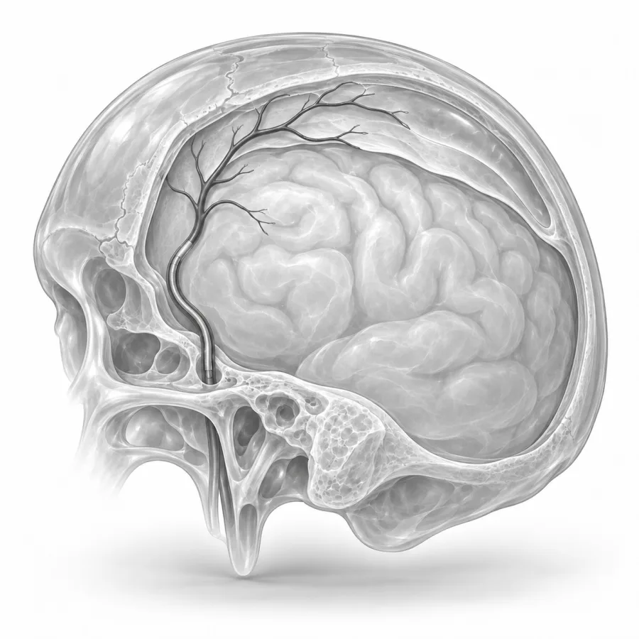
 On imaging
Side view of a cerebral angiogram, the map we use to find the middle meningeal artery.
Middle Meningeal Artery Embolization
Dr. Fabio Komlos walks through middle meningeal artery embolization the way he explains it in clinic, using the same images he uses to plan treatment.
Fabio Komlos, MDInterventional radiologist · 6:27Brain Bleeds to Fibroids: Same Results, Tiny Incision
2:25
After Embolization? Don't Make These 5 Mistakes
5:32
What is a chronic subdural hematoma?
The brain is covered by several thin layers. The toughest outer layer, just under the skull, is called the dura. A subdural hematoma is a collection of blood that forms between the dura and the brain. A chronic subdural hematoma (cSDH) builds up slowly, over weeks, often after a minor bump to the head that you may not even remember.
It is most common in people over 65. As we age, the brain shrinks a little and the small veins bridging to the dura stretch and tear more easily. Blood thinners, antiplatelet medicines such as aspirin, frequent falls and heavy alcohol use raise the risk.
Once a chronic subdural starts, the body forms a thin, inflamed lining around it, called a membrane, full of fragile new blood vessels. These vessels keep leaking small amounts of blood, so the collection can grow or come back even after it is drained. Many of these vessels are fed by the middle meningeal artery (MMA), an artery that runs along the inside of the skull and feeds the dura.
Symptoms and when to worry
- Headache that is new or getting worse
- Confusion, memory trouble or personality changes, which family members often notice first
- Weakness or numbness on one side of the body
- Trouble walking, balance problems or falls
- Trouble speaking or finding words
- Drowsiness or, in severe cases, seizures
How MMA embolization works
Embolization means blocking a blood vessel on purpose. In MMA embolization, we guide a thin catheter into the middle meningeal artery and fill its branches with a liquid embolic agent that hardens like soft glue, or with tiny particles. This cuts off the blood supply to the leaky membrane. With the leak stopped, the body can slowly absorb the old blood over the following weeks to months.
The dura has other sources of blood, so blocking the MMA does not harm the brain. The brain itself is fed by different arteries that are not touched.
What we seeWho it helps and who it may not
Good candidates
- After surgery to drain a chronic subdural, to lower the chance it comes back
- Smaller collections with mild symptoms, instead of surgery
- People with a collection that keeps coming back
- People who need to restart blood thinners soon
- People who are at high risk from surgery or anesthesia
May not be right if
- Severe or rapidly worsening symptoms from pressure on the brain, which need urgent surgical drainage first
- A new (acute) bleed from a serious injury
- Artery anatomy that connects the MMA to the arteries of the eye or brain in a way that cannot be made safe
- Severe allergy to contrast dye or serious kidney problems (can sometimes be managed)
Decisions are made together with a neurosurgeon. MMA embolization does not remove the blood that is already there, so it does not give fast relief from pressure on the brain. When a large collection is causing significant symptoms, surgery comes first, and embolization is often added a day or two later.
How it compares with the alternatives
| MMA embolization | Surgical drainage | Watchful waiting | |
|---|---|---|---|
| How it's done | Catheter through the wrist or groin | Burr holes or a small skull opening | Repeat CT scans |
| Anesthesia | Moderate sedation or general | General, sometimes local | None |
| Relieves pressure quickly | No, works over weeks | Yes | No |
| Chance of coming back | Low, especially when combined with surgery | About 1 in 10 or more with surgery alone | Collection may grow |
| Best for | Preventing recurrence; mild cases | Large collections with symptoms | Small collections with no symptoms |
Large studies published in 2024 found that adding MMA embolization to standard treatment, including surgery, cut the chance of the hematoma coming back or needing another operation roughly in half or more. Some small collections without symptoms are simply watched, sometimes after pausing blood thinners when safe to do so.
What happens, step by step
- 1Getting ready
An IV is placed, and you receive moderate sedation or general anesthesia so you can lie very still. We review your blood thinners with you and your neurosurgeon beforehand. See blood thinners.
- 2Reaching the artery
We numb the wrist or groin and place a small tube in the artery. Using X-ray, a catheter is guided up to the external carotid artery in the neck, and a very thin microcatheter into the middle meningeal artery.
- 3Blocking the branches
Contrast dye maps the artery and checks for connections to the eye or brain. We then slowly inject the liquid embolic agent or particles to fill the branches feeding the membrane, on one or both sides.
- 4Finishing
A final picture confirms the branches are closed. The tube is removed and the pinhole is sealed with a wrist band or closure device.
The part of this procedure I spend the most time on is the map. Before I inject anything, I look for small connections between the middle meningeal artery and the arteries to the eye and face, because protecting those is what keeps the procedure safe. I remind families that the blood collection does not disappear overnight. It shrinks over the next few months, and that slow change on the follow-up scans is what we are hoping to see.Fabio Komlos, MD, interventional radiologist · About Dr. Fabio Komlos
Recovery and aftercare
- First few hoursYou rest while nurses check the puncture site and do regular neurological checks: strength, speech and alertness.
- Night afterMost people stay 1 night for observation, often longer if embolization was done together with surgery.
- First 1–2 weeksSome people have a headache, jaw or temple ache for a few days. Avoid heavy lifting, straining and driving until your doctors clear you. Take steps to prevent falls at home.
- 2–6 weeksA follow-up CT scan checks the collection. It may look the same at first, which is expected.
- 3–6 monthsRepeat CT scans usually show the collection shrinking or gone. Your neurosurgeon will guide when blood thinners can be restarted, if needed.
Do not drive until your neurosurgeon clears you, especially if you have had a seizure or symptoms from the hematoma. Rules for driving after a seizure vary by state.
Results and risks
Most people have a steady shrinking of the collection over 3 to 6 months. When embolization is added to surgery, the chance of needing another operation drops substantially. A small number of people still need drainage later if the collection grows or symptoms get worse.
Possible risks
- Headache or pain in the jaw or temple for a few days
- Bruising or bleeding at the wrist or groin
- Stroke, if embolic material reaches the brain arteries (rare, about 1 in 100 or fewer)
- Vision loss, if material reaches the eye's artery through a hidden connection (very rare)
- Weakness of the face, from reduced blood flow to a facial nerve branch (rare, usually temporary)
- The hematoma growing despite treatment, needing surgery
- Reaction to contrast dye or kidney strain
Common questions
Is this brain surgery?
Does it hurt?
How soon will I feel better?
Can I take my blood thinner again?
Will I need more scans?
Will insurance cover it?
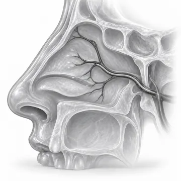
Epistaxis embolization
Stop severe or recurring nosebleeds by sealing the bleeding arteries from the inside.
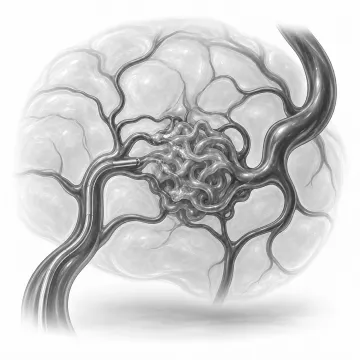
Arteriovenous malformation embolization
Close the abnormal artery-to-vein connections of an AVM through a pinhole, without surgery.

Blood thinners and your procedure
Which blood thinners may be paused before a procedure, typical hold times, and how bridging works.

Sedation and anesthesia
Local numbing, twilight sedation and anesthesia: what you feel and why you need a ride.
Written for patients by the Palo Alto Interventional team. General information only; your own plan depends on your health and imaging.
Talk to us about your options
Request a consultation online, or call 650-404-8445, Monday to Friday, 8 am to 5 pm. You can also ask your doctor to send a referral. We will review your imaging and help you decide whether a procedure makes sense.